
Computed Tomography (CT) – What You Need to Know
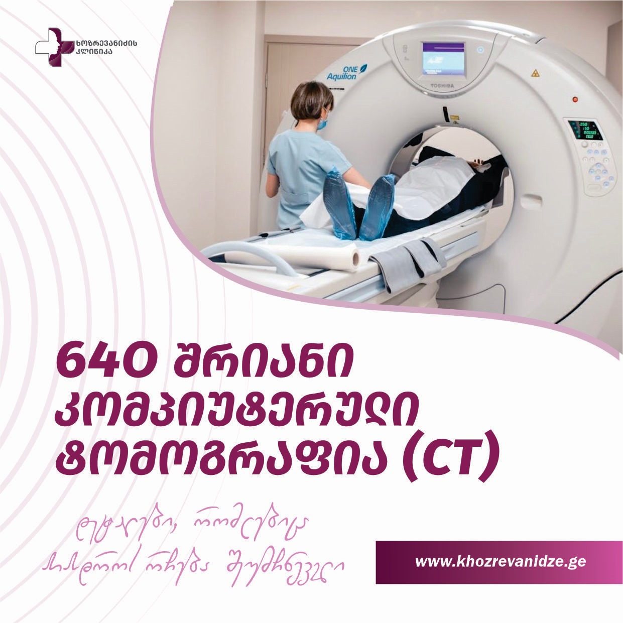
Computed Tomography (CT)is anon-invasive imaging methodthat performs thin-layer scanning of the body's internal structures. The obtained data are transformed by a special computer program intothree-dimensional (3D) images, allowing physicians to accurately assess organ structures, pathological changes, and disease spread.
The 640-slice CT scanner at Khozrevanidze Clinic representsworld-class technologycombining speed, precision, and patient safety. Its main advantages include: •Ultra-high resolution– clear visualization with 0.5 mm slices. •Minimal radiation– a special dose control system reduces radiation exposure, especially for children and the elderly. •High scanning speed– a complete organ can be scanned within seconds. •3D reconstruction– enables detailed assessment of organs or vascular systems in multiple planes and spatial projections. •Motion control– minimizes motion artifacts during heart and lung scans, crucial for cardiac diagnostics.
Main Clinical Applications of CT
CT is widely used in almost all medical fields: •Brain and skull– diagnosis of stroke, hemorrhage, tumors, and trauma. •Chest organs– detection of lung diseases (pneumonia, embolism, oncology), and evaluation of mediastinal structures. •Cardiac CT / CT Coronary Angiography– non-invasive evaluation of coronary arteries and calcium index. •Abdominal and pelvic organs– precise imaging of the liver, kidneys, pancreas, intestines, and reproductive organs. •Musculoskeletal system– detailed assessment of fractures, degenerative, and tumor processes. •CT Angiography– visualization of blood vessels throughout the body to detect aneurysms, stenoses, and thromboses.
Preparation
After scheduling, you may receive apreliminary phone consultationwith a doctor regarding preparation, depending on your individual needs.
Procedure
CT isfast and painless. The patient lies on the scanner table, and scanning lastsa few seconds to a few minutes, depending on the exam type. In some cases, acontrast agentis used to highlight vessels and tissue structures, improving diagnostic accuracy. The clinic maintainsstrict radiation dose control, and all procedures are performed under the supervision ofexperienced radiologists and technicians.
Results
After the scan, the images are processed with3D reconstruction software. The radiology team at Khozrevanidze Clinic provides adetailed report and conclusion, available inprinted and/or digital format(on CD or via patient portal).
Why Choose Khozrevanidze Clinic?
- Latest Toshiba Aquilion ONE 640-slice CT scanner • Experienced team of radiologists • Accurate, fast, and safe diagnostics • Individual approach to each patient • Procedures meeting international standards
Khozrevanidze Clinic – where precision means health.📞 0422 22 71 71 📍 Address: Batumi, Giorgi Brtskinvale St. 81
Frequently Asked Questions (FAQ)
Is CT painful?No. The scan is painless. With contrast, you may briefly feel warmth or a metallic taste.
How long does it take?Scanning takes1–5 minutes; the total visit usually lasts15–30 minutes.
Should I fast?For contrast studies – yes,4–6 hoursbefore. For non-contrast – usually not required.
I'm allergic to iodine. Can I still have a CT?Possibly, but a prior evaluation and premedication/alternative approach may be needed.
I have claustrophobia.CT isopen-type and fast; calming methods can be discussed with your doctor if needed.